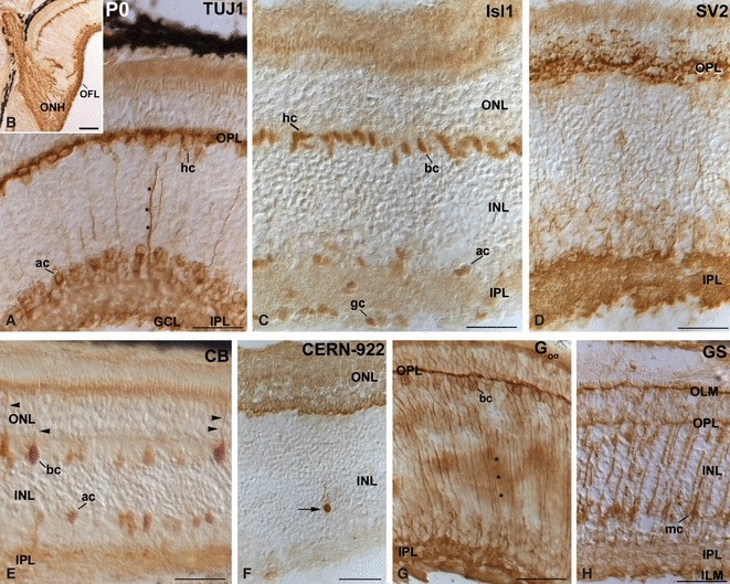
Fig. 4.

Patterns of expression of cell markers in the small-spotted catshark retina at hatching (P0). (A, B) TUJ1 immunoreactivity was detected in many horizontal and amacrine cells, in sparse ganglion cells, in fine processes oriented along the vitreo-scleral axis (asterisks), and in the OFL. Strong immunoreactive ganglion cell axons were also observed in the ONH (B). (C) Isl1 immunoreactivity was detected in the nuclei of subpopulations of horizontal, bipolar, amacrine and ganglion cells. (D) The IPL and OPL were highly immunoreactive to the SV2 antibody. Fine processes distributed throughout the INL appeared faintly immunostained. (E) Immunoreactivity against CB was mainly detected in bipolar and amacrine cells. Many bipolar cells exhibited apical processes that ran towards the OLM (arrowheads). Descending appendages from amacrine cells were also observed. The OPL and IPL appeared faintly immunostained. (F) Most of the photoreceptor cells were labelled with CERN-922 antibody. Occasional opsin-immunoreactive cells were observed in the INL (arrow). (G) Goα-immunopositive cell bodies were located in the scleral part of the INL. Fine long immunoreactive processes that reached the IPL (asterisks) were also immunolabelled. The IPL and OPL were immunoreactive. (H) GS staining was evident in cell somata located in the vitreal half of the INL, and in vitreal and scleral processes of radially oriented cells. The OLM, ILM and OPL also appeared stained. Scale bars: 25 μm (A, C–H); 100 μm (B). ac, amacrine cell; bc, bipolar cell; CB, calbindin; gc, ganglion cell; GCL, ganglion cell layer; hc, horizontal cell; ILM, inner limiting membrane; INL, inner nuclear layer; IPL, inner plexiform layer; mc, Müller cell; OFL, optic fibre layer; OLM, outer limiting membrane; ONH, optic nerve head; ONL, outer nuclear layer; OPL, outer plexiform layer.
