Fig. 5.

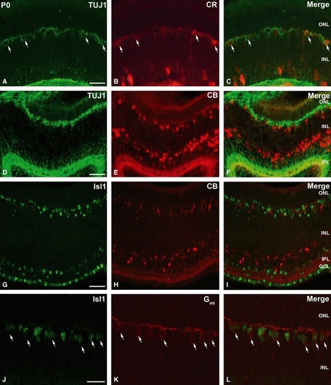
Photomicrographs of P0 small-spotted catshark retina to illustrate the colocalization of CR (green) and TUJ1 (red) (A–C), TUJ1 (green) and CB (red) (D–F), Isl1 (green) and CB (red) (G–I), and Goα (green) and Isl1 (red) (J–L). (A–C) Panels show localization of TUJ1 in CR-immunoreactive horizontal cells (arrows). (D–F) CB was not expressed in TUJ1-immunoreactive horizontal cells. (G–I) Panels illustrate the same section labelled for Isl1 and CB, where no colocalization was detected. (J–L) Goα-immunoreactive bipolar cells also expressed Isl1 (arrows). Scale bars: 25 μm (A–I); 15 μm (J–L). CB, calbindin; CR, calretinin; GCL, ganglion cell layer; INL, inner nuclear layer; IPL, inner plexiform layer; ONL, outer nuclear layer.
