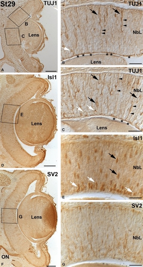
Fig. 7.

Early immunochemical markers in the embryonic small-spotted catshark retina at St29. The boxed areas in (A, D, F) are shown at higher magnification in (B, C, E, G). (A–C) Many TUJ1-positive somata were found in the central retina with their cell bodies located at varying distances from the vitreal surface (black arrows in B, C) and in the presumptive GCL (white arrows in B, C). Immunoreactive vitreo-scleral processes (arrowheads in B, C) and optic axons (asterisks in B, C) were distinguishable. (D, E) The number of Isl1-positive nuclei increased by this stage (D, arrows in E). Many of these immunoreactive nuclei were close to the inner surface of the neuroretina (white arrows in E). (F, G) Radial cell prolongations immunoreactive for SV2 were dispersed throughout the neuroepithelium by this stage, mainly in the central retina. Strong immunoreactive ganglion cell axons were observed in the optic nerve. Scale bars: 100 μm (A, D, F); 25 μm (B, C, E, G). NbL, neuroblastic layer; ON, optic nerve.
