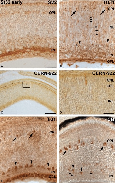
Fig. 9.

Neurochemical profiles in the embryonic central retina at St32-early. The boxed area in (C) is shown at higher magnification in (D). (A) SV2 antibody revealed the emergence of the OPL in the central retina. Furthermore, strong immunoreactivity was detected in the IPL. (B) The rounded TUJ1-immunoreactive somas, located close to the OPL, represented putative differentiating horizontal cells (arrows). Immunostaining was detected also in amacrine cells (large arrowheads), in descending cell prolongations (small arrowheads) located in the INL, IPL and OFL. (C, D) Scarce opsin-immunoreactive photoreceptor cells, showing morphological features of immaturity, were observed in the ONL. (E) Isl1 antibody recognized for the first time cell nuclei located in the outer region of the INL (arrows). Immunoreactivity was also found in most of the ganglion cells and in a subpopulation of amacrine cells located in the innermost region of the INL (arrowheads). (F) CB immunoproducts were found in somas located in the outer region of the INL (arrows), and in cell perikarya (large arrowheads) and their processes (small arrowheads) in the vitreal part of the INL. Scale bars: 25 μm (A, B, D–F); 100 μm (C). CB, calbindin; GCL, ganglion cell layer; INL, inner nuclear layer; IPL, inner plexiform layer; OFL, optic fibre layer; ONL, outer nuclear layer; OPL, outer plexiform layer.
