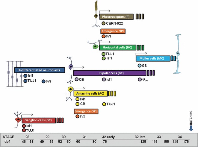
Fig. 11.

Schematic diagram of the onset of cell marker expression in the central retina for the different retinal cell types. The earliest detectable time of cell marker onset in a cell population is indicated as dots. Note that sometimes not all cells within a certain cell population are necessarily labelled, for instance Is1 amacrine cells or CB bipolar/amacrine cells. The time course of expression of different cell markers showed a vitreal to scleral progression of cell differentiation in the shark retina. CB, calbindin; GS, glutamine synthetase; OPL, outer plexiform layer
