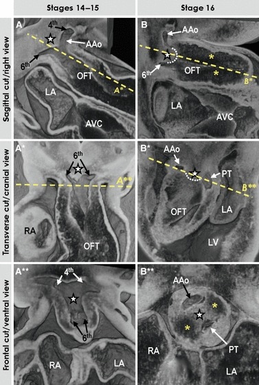
Fig. 4.

Three-dimensional (3D) morphology of the distal outflow tract, as assessed by high-resolution episcopic microscopy. (A–A**) Different views of the unseptated distal outflow tract at stages 14–15 with the dorsal wall between the 4th and 6th aortic arches slightly protruding intrapericardially (star), which leads to the appearance of the future extrapericardial ascending aorta (AAo). (B–B**) Different views through the septated distal outflow tract at stage 16. Note that progressive protrusion of the dorsal wall of the outflow tract intrapericardially leads to the formation of the so-called aortopulmonary septum (#, outlined by the dotted line in B, B*) separating the developing intrapericardial aortic channel from the future pulmonary trunk (PT). Yellow asterisks indicate endocardial cushions. See text for further description.
