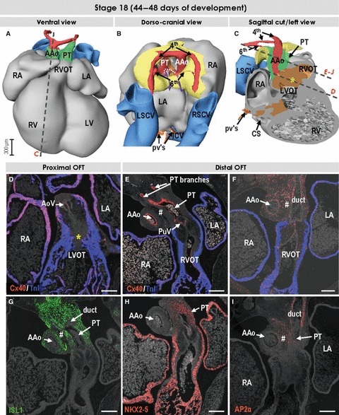
Fig. 7.

Three-dimensional (3D) and molecular analysis of the cardiac outflow tract at stage 18. (A–C) The myocardial outflow tract has further shortened, along with formation of the right ventricular infundibulum (RVOT). The intrapericardial ascending aorta and pulmonary trunk now possess their own discrete non-myocardial walls. The dotted line in (B) points to the proximal parts of the 6th arches, which now form the bifurcation of the pulmonary trunk. The left-sided 6th arch is persisting to become the arterial duct, while the right-sided 6th arch distal to the origin of the right pulmonary artery is regressing. Both 4th arches are of roughly equal size, while the third arches are no longer recognisable. (D) Section through the left ventricular outflow tract (LVOT) and developing aortic valve (AoV). The asterisks in (C) and (D) refer to the myocardialising mesenchyme between the ventricular outflow tracts. (E–J) Sections through the right ventricular outflow tract, developing pulmonary valve (PuV), pulmonary trunk and ascending aorta were incubated with antibodies as indicated. The tissue between great arteries (#) is ISL1-negative and expresses AP2α, which is also expressed in the facing walls of the arterial trunks. In contrast to the wall of the ascending aorta, the pulmonary truncal wall expressed transcription factor NKX2-5, reflecting the cardiogenic potential of these mesenchymal cells. Scale bars: 200 μm. For abbreviations, see previous figures.
