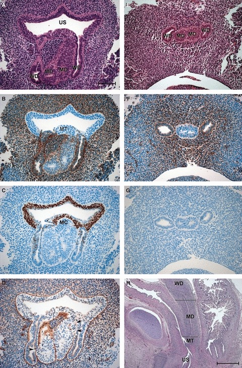
Fig. 1.

Sections of the vaginal anlagen (VA) of 8- and 10-week embryos. (A–D) Neighbouring transverse sections through the VA of an 8-week embryo at the level of the urogenital sinus. (A) HE staining; (B) vimentin; (C) p63; (D) laminin (arrowheads point to gaps in the basal lamina). (E–G) Neighbouring transverse sections through the VA of the same embryo at the level of the lower utero-vaginal canal. (E) HE staining; (F) vimentin; (G) p63. Bar: 100 μm. (H) Sagittal section through the VA of a 10-week fetus. The level of A–D is marked with the caudal line, whereas that of E–G is indicated by the cranial line. Bar: 1000 μm. MC, mesodermal cells; MD, Müllerian duct; MT, Müllerian tubercle; US, urogenital sinus; WD, Wolffian duct.
