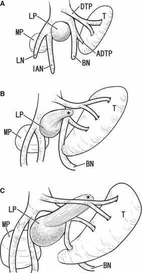
Fig. 9.

Schematic representation of a developmental change in the topographical relation between the lateral pterygoid muscle and buccal nerve. Viewed from the lateral side. The right-hand side of the figure corresponds to the anterior side of the head. In the early stage or at 6–7 weeks of gestation (panel A), the lateral pterygoid muscle anlage (LP) is restricted at a wedged position between the buccal nerve (BN) and the inferior alveolar nerve (IAN). The anlage of the temporalis muscle (T) is located superior to the lateral pterygoid muscle. In the intermediate stage (8–9 weeks; panel B), the buccal nerve changes direction at a branching site of the anterior deep temporal nerve (ADTP). The perspective upper head or the fetal anterior muscle slip of the lateral pterygoid muscle (black star) extends anterosuperiorly between the buccal nerve and the other deep temporal nerves (DTN). In the late stage (approximately 14–20 weeks; panel C), the upper head grows significantly and pushes the deep temporal nerve to the sphenoid bone. The buccal nerve may make an impression when the expanding temporalis muscle attacks the nerve (arrow in panel C). The actual difference in size of the muscles between panels is much more evident than it appears in the figures. LN, lingual nerve; MP, medial pterygoid muscle.
