
Figure 4. MR images of MRL/lpr and MRL/Mpj mice.
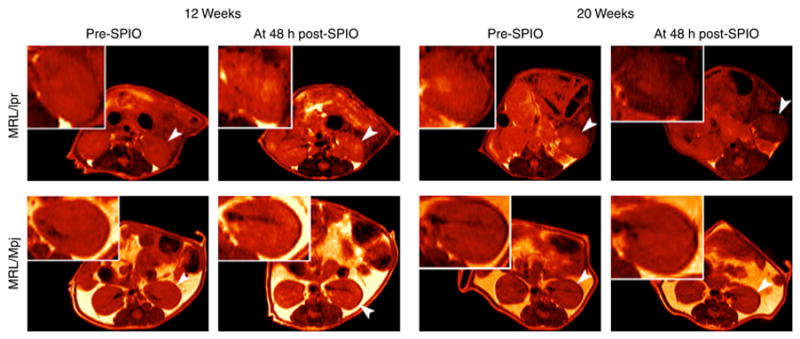
Representative abdominal MR images at time to echo (TE) 20 ms of 12 and 20 weeks old MRL/lpr and MRL/Mpj mice taken prior to (pre-SPIO) and 48 h following (post-SPIO) CR2-targeted SPIO injection. The MR images are colored red for better visual presentation. A reduction in intensity is observed for left kidney (arrowhead and insert) from pre- to post-SPIO injection for 20 weeks old MRL/lpr mouse. In this same MRL/lpr mouse, the right kidney, although not as dark as the left, is darker than the right kidney of the same mouse at pre-SPIO injection. Of note, T2-relaxation times (determined from MR images of kidneys generated from 16 different TE) are a more sensitive measure for detecting SPIO-induced MRI signal reduction than visual evaluations of MR images, all of which are obtained at a single TE.