
Figure 5. Non-invasive detection of glomerulonephritis progression.
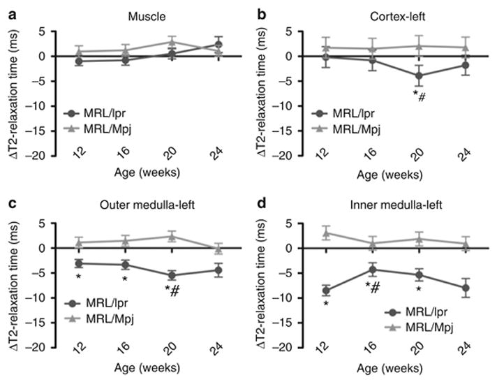
T2-relaxation times were measured prior to CR2-targeted SPIO injection and 48 h following injection. Changes in T2-relaxation times from pre-injected values ( T2-relaxation times in milliseconds (ms)) for (a) muscle, (b) left kidney cortex, (c) outer medulla and (d) inner medulla of MRL/lpr and control MRL/Mpj mice at 12, 16, 20 and 24 weeks are presented. The right kidneys of MRL/lpr and MRL/Mpj mice had similar values (data not shown, Supplementary Table S1). When ΔT2-relaxation times were compared between the two strains, the MRL/lpr mouse kidneys show significantly reduced ΔT2-relaxation times at 20 weeks (cortex) and 12, 16 and 20 weeks (outer and inner medullae) from those of age- and area-matched MRL/Mpj mouse kidneys (* p < 0.05). When ΔT2-relaxation times were compared between the ages within the same strain, the MRL/lpr mouse kidneys showed a significant decrease in ΔT2-relaxation time at 20 weeks for cortex and outer medulla, and at 16 weeks for inner medulla (# p < 0.05) when compared to earlier ages.