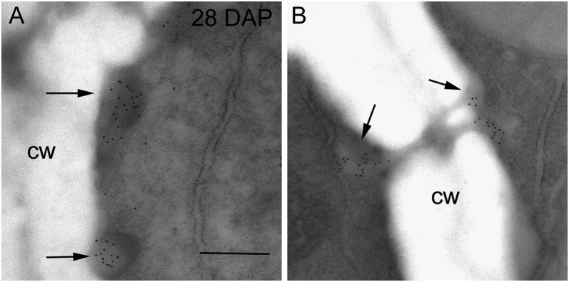
Figure 2.
Transmission electron micrographs of endosperm cells at 28 DAP labeled with (1→3)-β-d-glucan antibody. A, Deposits of callose were seen in electron-dense regions at the edges of subaleurone cells (arrows). B, In the starchy endosperm gold labeling was restricted to the callosic-rich plugs surrounding plasmodesmata (arrows). Bar = 0.5 μm. cw, Cell wall.

