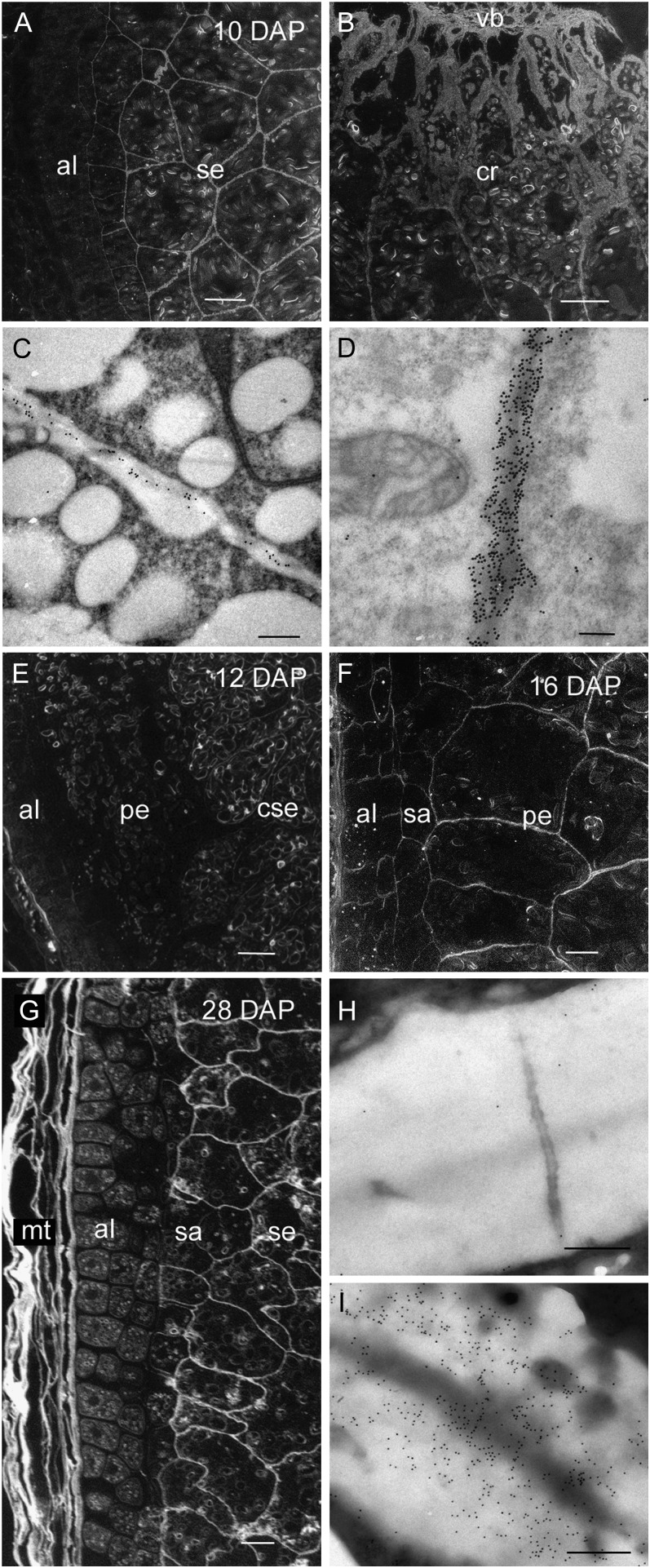
Figure 6.
Light and transmission electron micrographs of hetero-(1→4)-β-d-mannan labeling during endosperm differentiation from 10 to 28 DAP. A, Silver-enhanced antibody labeling at 10 DAP revealing little or no hetero-(1→4)-β-d-mannan in the aleurone cell walls whereas the starchy endosperm is well labeled. Bar = 100 μm. B, At 10 DAP the thicker starchy endosperm cell walls of the crease region are heavily labeled. Bar = 200 μm. C, Antibody labeling at 10 DAP revealing low levels of hetero-(1→4)-β-d-mannan in the aleurone cell walls. Bar = 0.5 μm. D, The cell walls of the starchy endosperm are heavily labeled at 10 DAP. Bar = 0.5 μm. E, Silver-enhanced antibody labeling at 12 DAP showing little labeling in the aleurone cell walls, no labeling in the walls of the peripheral starchy endosperm whereas hetero-(1→4)-β-d-mannan is present in the walls of the central starchy endosperm. Bar = 100 μm. F, By 16 DAP the walls of the peripheral starchy endosperm are labeled. Bar = 100 μm. G, At 28 DAP, silver-enhanced antibody labeling shows hetero-(1→4)-β-d-mannan is present in the walls of the maternal tissues, subaleurone, and starchy endosperm but absent in the thickened aleurone cell walls. Bar = 100 μm. H, Gold-labeled sections confirm hetero-(1→4)-β-d-mannan is absent in the aleurone cell walls but present in the starchy endosperm cell walls (I). Bars = 0.5 μm. al, Aleurone; cse, central starchy endosperm; cr crease region; mt, maternal tissues; pe, peripheral endosperm; sa, subaleurone; se, starchy endosperm; vb; vascular bundle.

