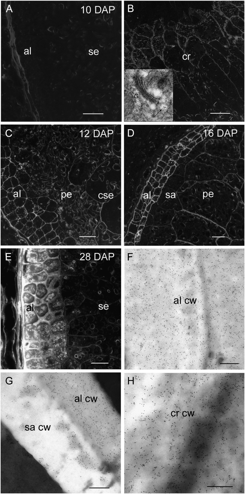
Figure 7.
Light and transmission electron micrographs of arabino-(1-4)-β-d-xylan labeling using the LM11 antibody during endosperm differentiation from 10 to 28 DAP. A, Silver-enhanced antibody labeling at 10 DAP reveals no arabino-(1-4)-β-d-xylan in aleurone or starchy endosperm cell walls. Bar = 200 μm. B, Arabino-(1-4)-β-d-xylan is present in the starchy endosperm cells walls of the crease region at 10 DAP. Bar = 200 μm. Inset, An electron micrograph of a gold-labeled Golgi apparatus (arrow) located within a cell at the crease. C, At 12 DAP silver-enhanced antibody labeling reveals arabino-(1-4)-β-d-xylan is now present in the aleurone and central starchy endosperm cell walls but absent in the peripheral starchy endosperm. Bar = 100 μm. D, At 16 DAP the cell walls of the peripheral starchy endosperm are labeled with the arabino-(1-4)-β-d-xylan antibody. Bar = 100 μm. E, At 28 DAP the aleurone cell walls are intensely labeled with the arabino-(1-4)-β-d-xylan antibody whereas labeling in the starchy endosperm cell walls is barely detectable. Bar = 100 μm. F, At 28 DAP electron micrographs of antibody-labeled sections show intense gold labeling in the aleurone cell wall. Bar = 0.5 μm. G, A heavily labeled aleurone cell wall abutting a subaleurone cell wall that shows patchy gold labeling with the arabino-(1-4)-β-d-xylan Ab. Bar = 0.5 μm. H, At 28 DAP the cell walls in the crease region continue to be strongly gold labeled. Bar = 0.5 μm. al, Aleurone; cw, cell wall; cse, central starchy endosperm; cr, crease; pe, peripheral endosperm; sa, subaleurone; se, starchy endosperm.

