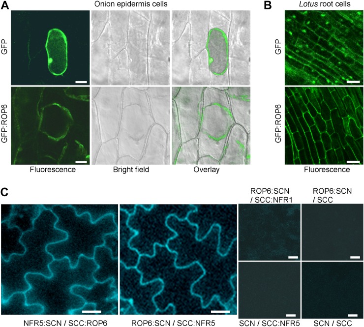
Figure 3.
Subcellular localization of ROP6 and its interaction with NFR5 in planta. A, Localization of GFP-tagged ROP6 in onion cells. Plasmids expressing GFP alone or GFP:ROP6 were delivered to the onion epidermal cells via particle bombardment. Onion epidermal cells expressing GFP:ROP6 were treated with 4% NaCl for 5 min to induce plasmolysis before imaging. Bars = 50 μm. B, Localization of GFP-tagged ROP6 in L. japonicus hairy roots. GFP alone served as a control. Bars = 20 μm. C, Interaction of ROP6 and NFR5 in planta. Cyan fluorescent protein (CFP) was split into N and C termini, which were fused to NFR5 and ROP6, respectively. The two constructs (NFR5:SCN and SCC:ROP6) were used to cotransfect tobacco leaves via Agrobacterium-mediated transient expression. The two fusion proteins were swapped with their CFP tags, and the resulting constructs (ROP6:SCN and SCC:NFR5) were coexpressed. Constructs SCC:NFR1 and CFP tags (SCN and SCC) were used to make combinations for coexpression and served as negative controls. Bars = 50 µm.

