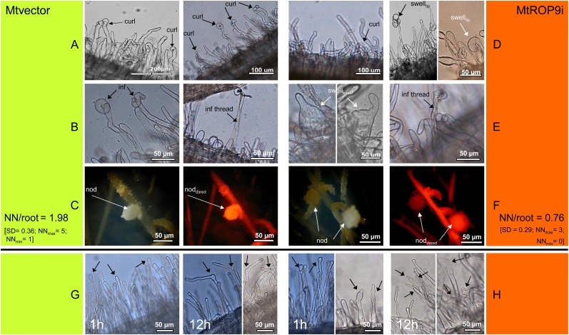
Figure 5.
Microscopic characterization of S. meliloti infection (A–F) and of root hairs treated with S. meliloti NF for 1 h (left panels) and 12 h (right panels; G and H) in MtROP9i and Mtvector transgenic roots. A, Rhizobia attach to root hairs (curl, curling). B, Early infection (inf, transcellular infection; inf thread, formation of infection thread). C, Early nodule (nod) formation (noddsred, DsRED-positive nodule). D, Rhizobia attach to root hairs (swelltip, abnormal swelling of tips). E, Left, interrupted infection (swellbasal, abnormal basal swelling of root hairs); right, successful infection. F, Early nodule formation. Text at C and F is as follows: number of nodules (NN) per single root at 21 dpi; NNmax, maximum number of nodules found in one root; NNmin, minimum number of nodules found in one root. G, Arrows indicate root hair tip swelling, branching, and reinitiation of polar growth. H, Arrows indicate progressed (tip) swelling, spontaneous constriction, and branching of root hairs. [See online article for color version of this figure.]

