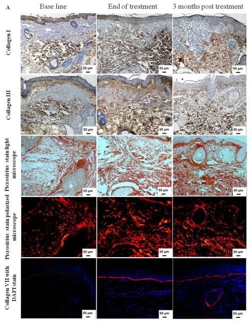
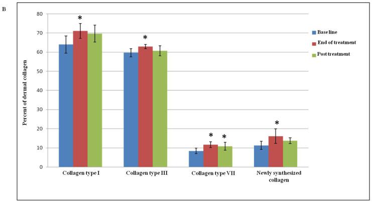
Figure 2.
(A) Increase in dermal collagen content after Er:YAG 2940 nm laser mini-peels. Immunohistochemical staining of skin tissues for collagen types I and III (1st and 2nd rows, respectively) showing an increase in collagen content. Shown in the 3rd and 4th rows are representative examples of skin tissues stained with picrosirius red viewed under bright field (3rd row) and polarized field (4th row). Bright field captures total collagen content while polarized light showed yellow to orange birefringence reflecting newly synthesized collagen in yellow to orange and total collagen in red. An increase in collagen type VII expression (red; 5th row) was observed after Er:YAG mini-peels compared to base line, and then decreased 3 months post treatment. Nuclei stained in blue with DAPI (Immunohistochemical and Picrosirius red; X 200); (B) Percent of dermis occupied by collagen levels. Data showed a statistically significant increase in both collagen types I, III and VII, as well as newly synthesized collagen at the end of Er:YAG laser mini-peels (*,p≤ 0.05).


