Figure 3.

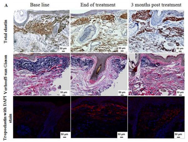
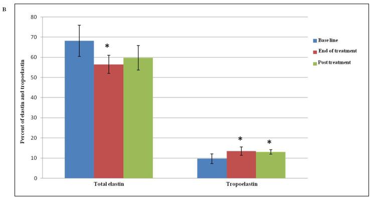
(A) Dermal elastin in response to Er:YAG laser mini-peels. Tissues were immunostained for total elastin (1st row) and Verhoeff-van Gieson stain (2nd row), showing a decrease in elastic fibers content. Immunofluorescence staining for tropoelastin (3rd row) shows increased deposition of newly synthesized tropoelastin in dermis (red), the sections were counterstained blue for nuclei with DAPI (Immunohistochemical and Verhoeff-van Gieson; X 200); (B) Percent of dermis occupied by elastin and tropoelastin showing significant changes after treatment. The values are mean ± SD (*, p ≤ 0.05).

