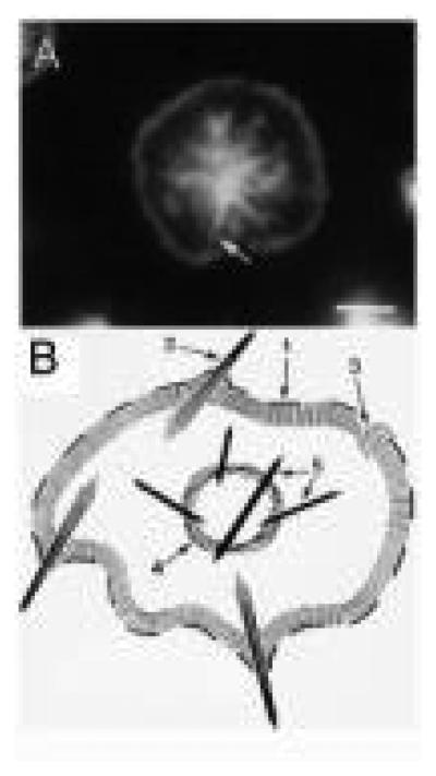
FIG. 3.
Actin structures by fluorescence. (A) An example of a platelet 15 min after spreading on glass imaged by fluorescence microscopy of F-actin as stained with phalloidin. Arrow indicates the position of a former filopodia. (B) Diagram of the actin structures in the spread platelets: (1) leading edge of the lamellipodium; (2) filopodia; (3) lamellipodium; (4) contractile ring; (5) stress fibers. (Reproduced from Bearer, 1995, by permission.)

