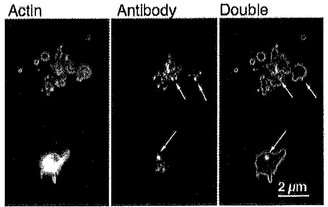
Figure 5. αArp2 antibody is detected in the cytoplasm after permeabilization.
Representative examples of platelets permeabllized, loaded with αArp2, activated on glass, and fixed as in Figure 3. After fixation, platelets were stained with Cy3-labeled secondary antibody to determine whether the primary antibody gained access to the cytoplasm during permeabilization. Staining for αArp2 (red) and actin filaments with FITC–phalloidln (green) demonstrates diffuse speckling in the cytoplasm with some larger aggregates of antibody (arrows).

