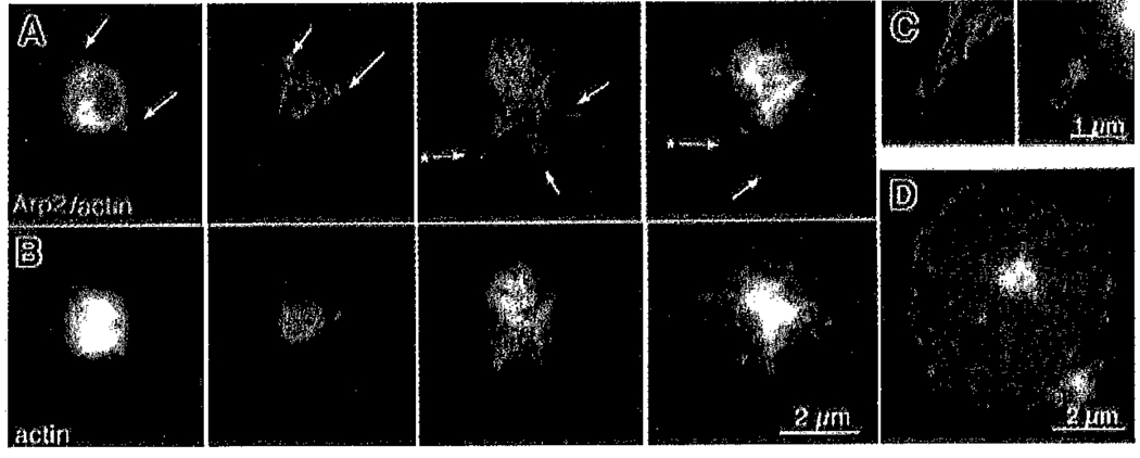
Figure 7. Gallery of platelets at early stages of shape change showing localization of Arp2/3 at the cortex and at the tips and roots of filopodia.
Representative double images of platelets fixed in the presence of detergent as in Figure 6A–B. (A) Four different platelets fixed at progressively later stages of activation from left to right and imaged for both F-actin (FITC–phalloidin, green) and Arp2/3 (Cy3, red). (B) Corresponding images of the same 4 platelets showing only the actin channel. (C) Higher magnification of the 2 filopodia indicated in panel A by an asterisk. (D) Fully spread platelet at the same magnification as the other platelets in this figure for size comparison.

