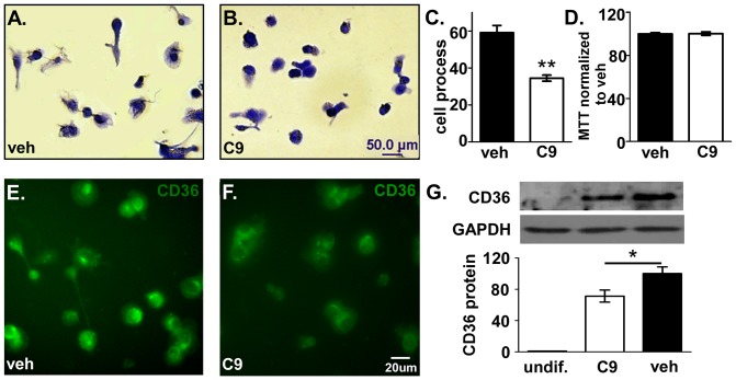
Figure 3. Inhibition of differentiation of HL-60 cells by CHEC-9.
HL-60 cells were treated with 50 nM CHEC-9 or TBS vehicle for 4days with stimulation by PMA. HL-60 cells adhere to plate surface, flatten out and grow processes (A). CHEC-9 treatment decreased the number of cells with process (B). Quantification of Coommassie blue staining (C). CHEC-9 did not affect viability of the cells (D). Immunostaining for CD36 showed that the fluorescent intensity of CHEC-9 group was decreased compared with vehicle group (E, F). This observation was supported by western analysis (G). Quantification was by normalization to GAPDH bands. (* p<0.05, ** p<0.005).

