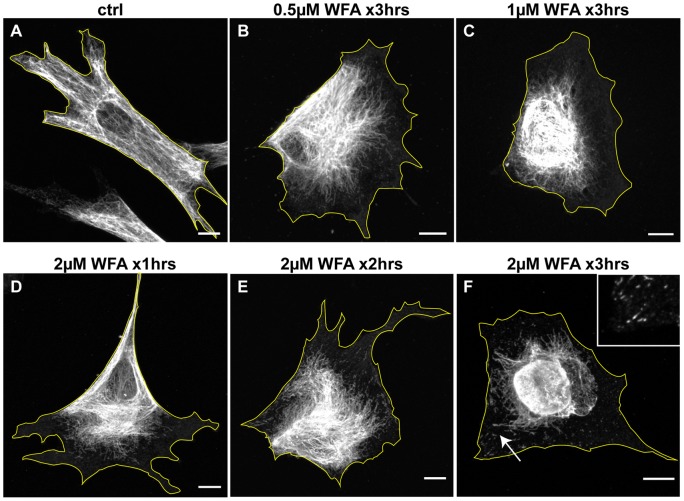
Figure 1. WFA treatment alters the subcellular organization of VIF.
BJ-5ta cells were treated for 3 hrs (A–C and F) with DMSO alone (A), 0.5 μM WFA (B), 1 μM WFA (C), and 2 μM WFA (F). In addition, cells treated with 2 μM WFA are depicted after 1 hr (D) and 2 hrs (E) which show that the changes in VIF organization take place gradually. Cells were fixed and processed for immunofluorescence with vimentin antibodies. Arrow: a region depicted at higher magnification in the inset, showing non-filamentous vimentin particles and short IF or squiggles. Scale bars =10 μm.

