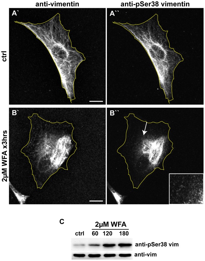
Figure 4. WFA treatment induces an increase in the phosphorylation of vimentin serine-38.
BJ-5ta cells were treated for 3 hrs with DMSO (A) or 2 μM (B) WFA, then fixed and double labeled with vimentin (A′ and B′) and pSer38 vimentin (A′′ and B′′) antibodies. Scale bars =10 μm. Arrow: a region depicted at higher magnification in the inset showing vimentin particles stained with pSer38 vimentin antibody. (C) Whole cell lysates of cells treated with DMSO (ctrl) or 2 μM WFA for 60 min, 120 min, and 180 min, were separated by SDS-PAGE and stained with anti-vimentin and anti-vimentin pSer38 vimentin antibodies.

