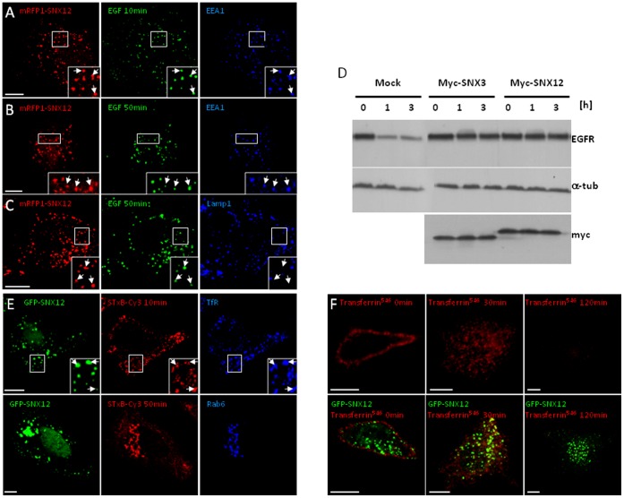
Figure 2. SNX12 overexpression inhibits EGFR transport and degradation without affecting retrograde and recycling transport routes.
(A-C) After cell surface binding, EGF-biotin coupled to streptavidinAlexaFluor488 was endocytosed for 10 min (A) or 50 min (B-C) at 37°C in HeLa cells expressing mRFP1-SNX12. Cells were labeled with anti-EEA1 (A) or Lamp1 (B-C) antibodies and analyzed by triple channel fluorescence. (D) HeLa cells expressing myc-SNX12 or myc-SNX3 or mock-treated were incubated with EGF for the indicated time periods. Cell lysates (100 µg) were analyzed by SDS gel electrophoresis and western blotting with antibodies against EGFR, α-tubulin (a-tub) or myc. (E) After cell surface binding, Shiga toxin B-subunit conjugated to Cy3 was internalized for 10 min or 50 min at 37°C into HeLa cells expressing GFP-SNX12. Cells were labeled with anti-transferrin receptor (TfR) or Rab6 antibodies and analyzed by triple channel fluorescence. (F) After cell surface binding (0 min), transferrin conjugated to AlexaFluor546 (transferrin546) was internalized for 30 min or 120 min at 37°C into control cells (upper panels) or cells expressing GFP-SNX12 (lower panels). Cells were then analyzed by fluorescence. (A-C and E-F) Scale bar indicates 10 µm.

