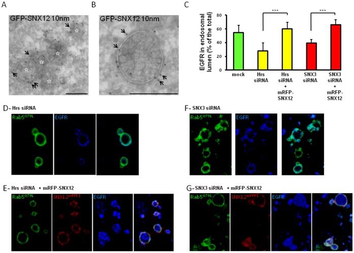
Figure 4. SNX12 overexpression induces an accumulation of MVB-like structures and rescues the intralumenal vesicles that incorporate the EGF receptor.
(A-B) HeLa cells expressing GFP-SNX12 were fixed and processed for electron microscopy. Cryosections were labeled with antibodies against GFP followed by protein A-gold 10 nm (arrows). Panel A shows a cluster of several multivesicular endosomes, each being labeled with a star, and panel B shows a high magnification view of an individual multivesicular endosome. Scale bar indicates 250 nm. (C) Hrs (D-E) or SNX3 (F-G) was knocked down and mRFP-SNX12 (red) was overexpressed (E-G) or not (D-F). In each condition, HeLa cells were also transfected with GFP-Rab5Q79L (green) during the last 24 h. After cell surface binding, EGF was internalized for 15 min at 37°C. Cells were processed for immunofluorescence with anti-EGFR antibodies (blue). The relative amount of EGFR in the lumen of endosome was quantified as previously described [25]. Results are expressed as the percentage of the total amount of EGFR. Each condition is the mean of at least three independent experiments; standard errors are indicated and results were analyzed by paired t test (***, p<0,001). (D-G) Representative pictures of experiments quantified in (C). GFP-Rab5Q79L enlarged endosomes in each condition showed the EGFR localization after Hrs (D-E) or SNX3 (F-G) was knocked down and mRFP-SNX12 (red) was overexpressed (E-G) or not (D-F).

