
Figure 4.
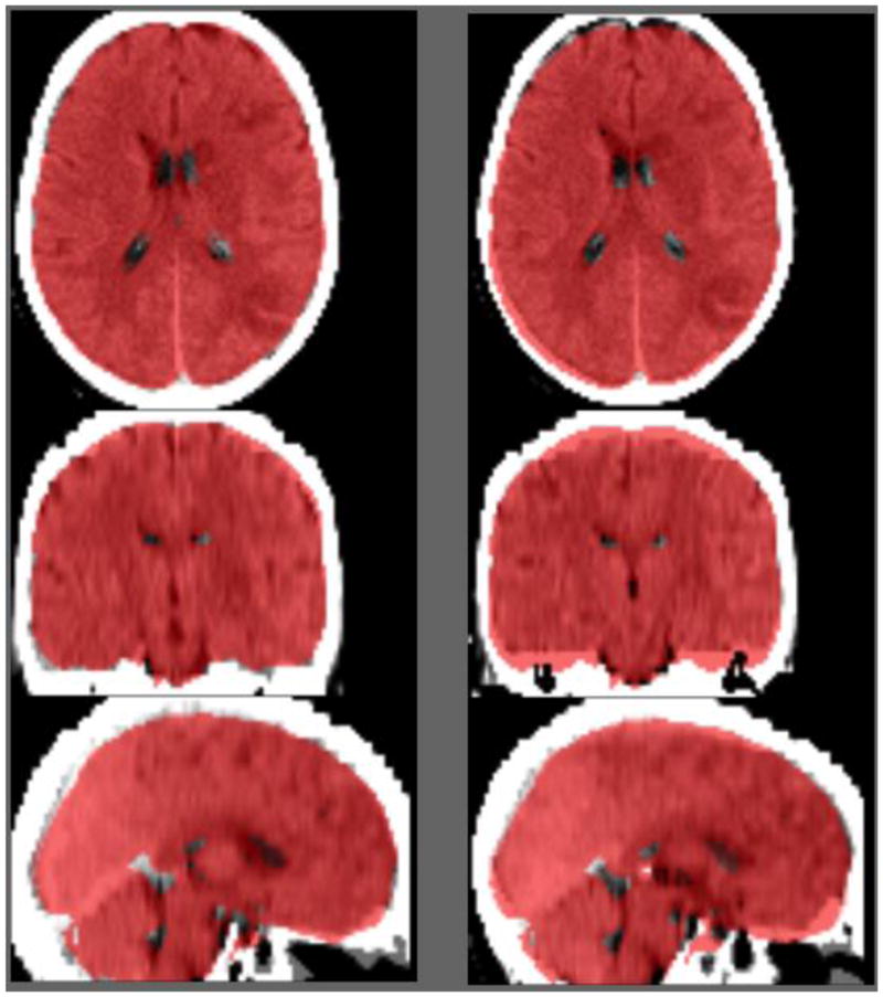
Example trial in our two-alternative forced choice trial. On each trial the observer examined the normalization of an acute stroke patient’s CT scan using two different normalization methods. The MNI brain mask (as distributed with FSL) is displayed overlayed on top of each image (red). The task is to report whether the left or right normalization was more successful. In this example, the normalization of the image using our new template is shown on the left and the performance of our ABLe clone is shown on the right.