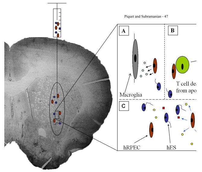
Figure 1. Schematic diagram of a grafting scenario of hRPEC cografted with hES grafts.
This diagram depicts hRPEC cografted with hES using stereotactic injection of cell suspension into the striatum. Enlarged schematic of cograft shows possible mechanisms of hRPEC (brown ovoid cells) providing localized immunosuppressed microenvironment that prevent deleterious host immune responses and promote growth and differentiation of hES (blue cells with neurite extensions). A. Possible role of hRPEC secreting immune modulating factors, thus suppressing microglial activation. B. hRPECs inducing apoptosis of cytotoxic T cells, thus decreasing the host cell mediated immune response. C. hRPECs could possibly provide various growth factors (yellow circles and red squares) to promote the growth and differentiation of the hES cells and to provide anti-teratoma effects.

