
Figure 1. Isx1 pharmacologically targets and regulates myocardial cells in vivo.
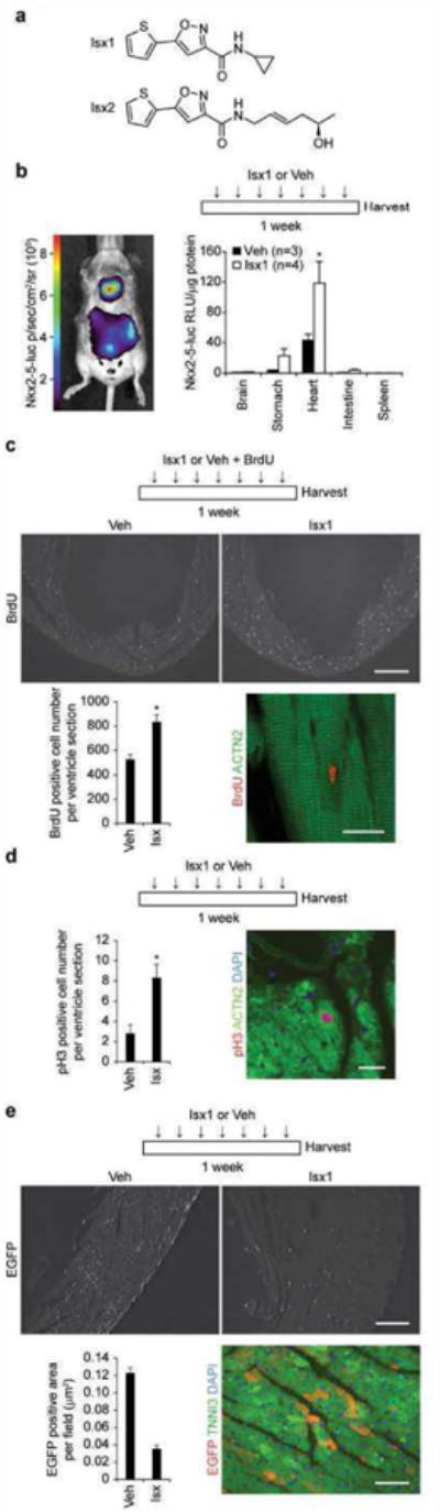
(a) Chemical structures of Isx1 (N-cyclopropyl-5-(thiophen-2-yl)isoxazole-3-carboxamide) and Isx2 ((R,E)-N-(5-hydroxyhex-2-enyl)-5-(thiophen-2-yl)isoxazole-3-carboxamide) compounds. (b) Time map of Isx1 in vivo treatment paradigm. Bioluminescence imaging of Nkx2-5-luc-BAC mouse demonstrating cardiac and stomach (a known extra-cardiac target) restricted luciferase activity after Isx injection in vivo. Quantitative evaluation of Isx-induced Nkx2-5-luc-BAC activity normalized per μg protein in tissue extracts from reporter mice injected ip once daily for 7 days with cyclodextrin (Veh, n=3) or Isx1 at 16 mg/kg (n=4). (c) Time map of Isx1 or Veh plus BrdU in vivo treatment paradigm. BrdU immunohistochemistry in uninjured adult mouse heart injected with vehicle or Isx (top panels, scale bar = 500μm). Quantitative evaluation of BrdU+ cells per ventricle section (9 sections from 3 animals were counted for each group, * = p<0.005). Immunohistochemistry localizing a BrdU+ nucleus (red) to an ACTN2+ cardiomyocyte (green) in an uninjured adult mouse injected with Isx (bottom right panel, Scale bar = 20μm). (d) Time map of Isx1 in vivo treatment paradigm. Quantitative evaluation of pH3+ cells per ventricle section in uninjured adult mice injected with Veh or Isx1 for 7 days (6 sections from 3 animals were counted for each group, * = p<0.05). Phospho-histone H3+ nucleus (red) localizing to an ACTN2+ cardiomyocyte (green), co-stained with DAPI (blue), in an uninjured adult mouse injected with Isx (right hand panel, Scale bar = 30μm). (e) Time map of Isx1 in vivo treatment paradigm. EGFP immunohistochemistry in uninjured adult mouse heart injected with Veh or Isx1 (top panels, scale bar = 100μm). Quantitative evaluation of EGFP+ area per field (18 fields per group) in uninjured adult mice treated with Veh or Isx1 for 7 days. EGFP+ (red) immunohistochemistry localized to TNNI3+ (green) cardiomyocytes, co-stained with DAPI (blue) (bottom right panel, scale bar = 30μm).