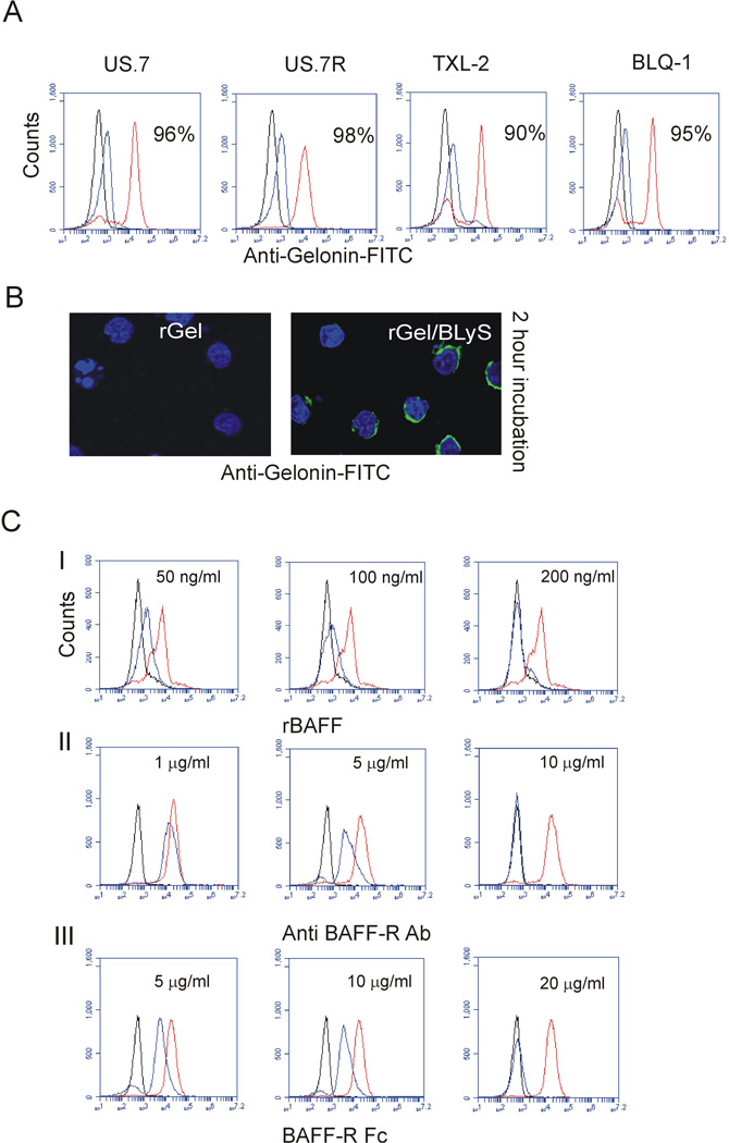
Figure 1. rGel/BLyS binds to human ALL cells.
(A) FACS analysis using anti-Gelonin antibodies for rGel (black) or cell surface rGel/BLyS (blue) and total rGel/BLyS (red) after incubation with 400 nM rGel/BLyS. Percentages indicate positivity for total rGel/BLyS (B) Immunohistochemistry for rGel or rGel/BLyS in US.7 cells. Note the apparent signal of rGel/BLyS as a perinuclear ring, due to the relatively large nucleus and small volume of cytoplasm in these cells. Green, Gelonin antibodies, blue, DAPI counter-stain for the nucleus. Images were captured using a Leica DIC analyzer, 200x 1.4–0.7, oil. (C) Detection of rGel or rGel/BLyS with anti-Gelonin antibody after pre-incubation of US.7 cells with the indicated concentrations of (I) recombinant human BAFF or (II) BAFF-R antibody; both then followed by incubation with 100 nM rGel/BLyS or (III) when BAFF-R Fc was added together with rGel/BLyS. (n=3). Controls; isotype (black) and positive control (no treatment, red); treated cells (blue).

