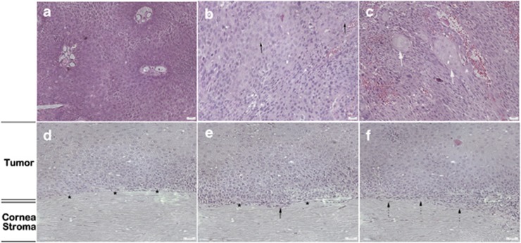
Figure 4.
Histopathological analysis of OSSN by H&E staining. (a) Enlarged and irregular squamous epithelial cells with abnormal density of cells (CIN). (b) Binucleated, actively mitotic dysmorphic cells (black arrows) and cells with increased nuclear/cytoplasm ratio were observed in patients diagnosed as carcinoma in situ. (c) Heteromorphic abnormal epithelial cells with partially formed carcinoma cell nests in OSSC were revealed (thick white arrows). The abnormal squamous epithelial cells (d) did not invade the Bowman's layer (black stars) in three cases diagnosed as CIN; (e) invaded but did not penetrate the Bowman's layer (black stars) in one case diagnosed as carcinoma in situ (black arrow); and (f) invaded beneath the Bowman's layer and into anterior stroma in the other case diagnosed as OSSC (black dashed arrows).

