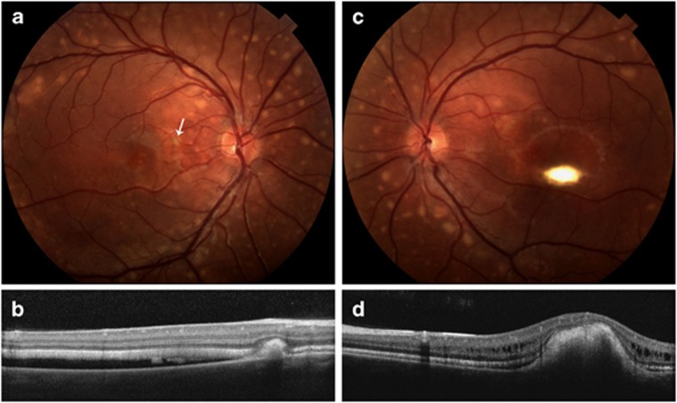
Figure 3.
(II-3) Fundus photographs (a, c) of both eyes significant for many small, scattered vitelliform lesions in the macula and larger vitelliform lesions along the arcades and in the periphery. Additionally, there are macular vitelliform lesions in both eyes with serous retinal fluid in the macula of the right eye. Optical coherence tomography images (b, d) through the macular and peripheral lesions are also shown. The peripheral lesion in the right eye (a, b) is located nasal to the macula (white arrow upper left panel), and the retina is also separated from the RPE. The scan through the macular lesion (d) shows elevation of the macular area with intraretinal fluid.

