Figure 1.
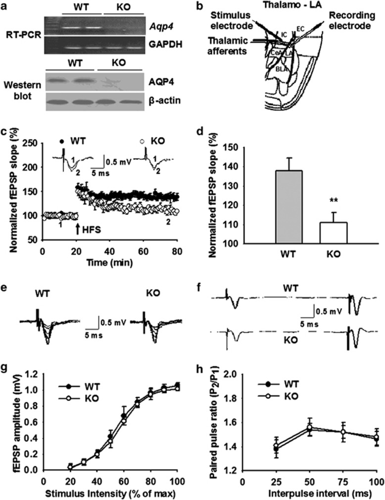
Aquaporin-4 (AQP4) deficiency impairs long-term potentiation (LTP) in the thalamo-LA pathway with no effect on basal synaptic transmission. (a) Expression of AQP4 in the LA from wild-type (WT) (AQP4+/+) and knockout (KO) (AQP4−/−) mice. Reverse transcriptase-polymerase chain reaction (RT-PCR) (upper) and western blot analysis (lower) revealed that the expression of Aqp4 mRNA and AQP4 protein was readily detected in LA of WT mice (n=3) but not KO mice (n=3). Glyceraldehyde 3-phosphate dehydrogenase (GAPDH) and β-actin were used as internal control. (b) Schematic representation of an amygdala slice showed location of recording and stimulation electrodes in the thalamo-LA pathway. LA, lateral amygdala; BLA, basolateral amygdala; CeA, central amygdala; IC, internal capsule; EC, external capsule. (c) Time course of the field excitatory postsynaptic potential (fEPSP) evoked by stimulation of thalamic inputs recorded in amygdala slices from WT (n=11 slices from 6 mice) and KO mice (n=13 slices from 7 mice). (Inset) Schematic representation of fEPSP recorded in individual slices before (1) and 60 min after (2) the LTP-inducing stimulation in either WT (left) or KO (right) mice. (d) The histogram showed the level of LTP 60 min after high-frequency stimulation (HFS) (five trains at 100 Hz for 1 s with 90 s interval between trains) in the thalamo-LA pathway in WT and KO mice. Each point was the normalized mean±SEM of slices. **P<0.01 vs WT. (e) Typically superimposed fEPSP recorded in the thalamo-LA pathway in WT (left) and KO (right) mice with gradually increased stimulation intensity. (f) Typical fEPSP recorded in the thalamo-LA pathway from individual experiment at 50 ms interpulse interval before HFS stimulation. (g) Input–output curves in the thalamo-LA pathway illustrating the relationship between the stimulation intensity and evoked response for fEPSP recorded in brain slices from WT (n=10 slices from 5 mice) and KO mice (n=11 slices from 6 mice). No significant differences were observed between the two genotypes. (h) Paired-pulse facilitation in the thalamo-LA pathway was measured by varying the intervals (25, 50, 75, and 100 ms) between pairs of stimuli before HFS stimulation. No significant differences were observed between WT (n=10 slices from 5 mice) and KO mice (n=11 slices from 6 mice).

