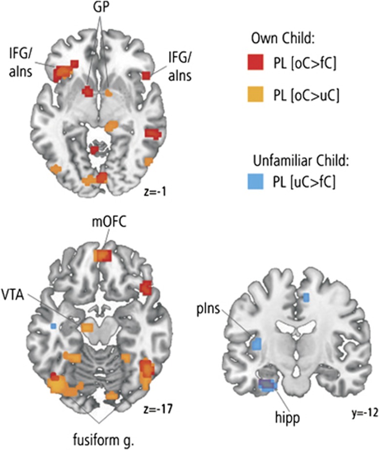
Figure 1.
Activation in response to own child pictures. Increased activations in response to oC>fC pictures under PL (red) were observed in the left GP, the mOFC, the left hippocampus, and the bilateral inferior frontal gyrus/anterior insula and fusiform gyrus. In response to oC>uC pictures under PL (yellow), a similar pattern of activations was observed including the right GP, mOFC, bilateral inferior frontal gyrus/anterior insula and fusiform gyrus, as well as the left VTA. The comparison of uC>fC pictures under PL (blue) revealed activations in the left posterior insula and the left hippocampus. Activated clusters are significant at puncorr<0.001 with a cluster extent threshold of k⩾7 voxels. aIns, anterior insula; fC, familiar child; hipp, hippocampus; IFG, inferior frontal gyrus; mOFC, medial orbitofrontal cortex; oC, own child; PL, placebo; pIns, posterior insula; uC, unfamiliar child; VTA, ventral tegmental area.

