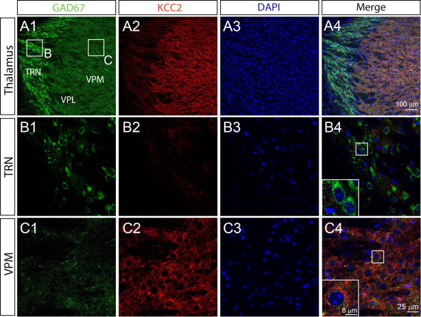
Figure 3.
Thalamic nucleus-specific expression of KCC2 A, Expression of GAD67 (A1) and KCC2 (A2) from the same section of mouse thalamus detected by immunohistochemistry. Cell nuclei were labeled by DAPI (A3). An overlay of the three channels is shown in A4. B, Higher magnification view of the immunoreactive signal of GAD67 (B1) and KCC2 (B2) for the area indicated in A1, showing KCC2 is not detectable in TRN neurons (B4). C, Higher magnification view of the immunoreactive signal for GAD67 (C1) and KCC2 (C2) for the area indicated in A1, showing KCC2 is strongly expressed in VPM neurons (C4). B3 and C3 show DAPI staining for TRN and VPM, respectively.

