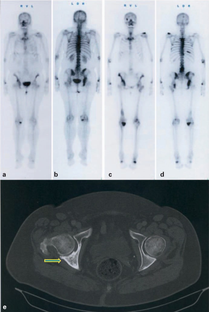
Fig. 2.
A 68-year-old female patient with breast cancer. Whole-body bone scan 3 h after intravenous injection of 656 MBq 99mTc-DPD, in anterior (a) and posterior view (b). Last post-therapy whole-body scan (c, d) 23 h after intravenous injection of 3.2 GBq 153Sm-EDTMP. Mild progressive disease after a cumulative activity of 17.0 GBq 153Sm-EDTMP; cancer antigen (Ca) 15–3 was increased from 65.5 U/ml (staging scan) to 175 U/ml 15 months later. (e) Computed tomography of the pelvis after the third 153Sm-EDTMP and continuous bisphosphonate therapy, showing calcification of a large, mainly lytic lesion of the pelvis (arrow).

