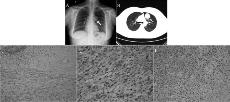
Fig. 1.
A 3.6 × 3.5 cm mass near the left pulmonary hilus (arrow); B Same mass in computed tomography image 4 months later (arrow); C Vaginal leiomyosarcoma containing interlacing fascicles of spindle cells, many with markedly pleomorphic nuclei, H&E ×200; D Vaginal leiomyosarcoma cells showing nuclear pleomorphism with bizarre nuclei and mitotic activity, H&E ×400; E Metastatic breast leiomyosarcoma cells with nucleic mitosis and necrosis, H&E ×200.

