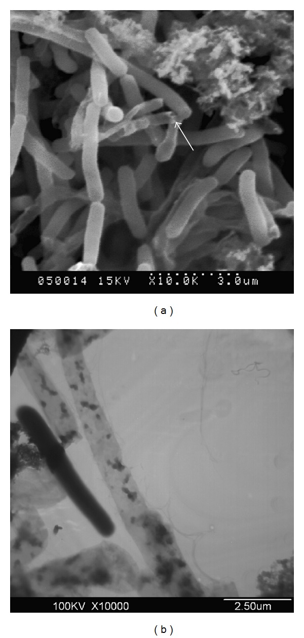
Figure 3.

Electron microscopy analyses of F. columnare G4cpN22 ghosts. (a) Arrow showed the transmembrane lysis tunnel located mainly at the cell poles via SEM. (b) Loss of cytoplasmic materials of F. columnare G4cpN22 ghosts was shown by TEM. The lysed cells showed uneven and low electron density and retained the basic cell morphology of the bacterial cells, while the unlysed cells showed even and high electron density in an integral cell structure.
