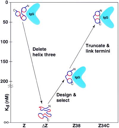
Figure 1.
Protein evolution in vitro. Minimization of the three-helix bundle Z domain into a two-helix bundle that maintains equivalent function with half the size. Z, natural three-helix domain. ΔZ, starting 38-residue truncated peptide. Z38, selected two-helix binding domain. Z34C, further truncated 34-residue peptide with engineered disulfide bond. Blue, hydrophilic amino acids; red, hydrophobic amino acids; green, disulfide bond.

