
Figure 1.
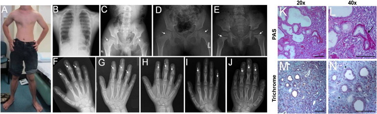
Clinical and Radiological Manifestations of MSS-Affected Individuals
(A and B) Photograph and thorax X-rays of affected individual FI1 at 17 years of age showing narrow chest.
(C–E) Hip X-rays of affected individuals FI1 (17 years), FIII1 (9 years), and FIII2 (5 years) showing flattened femoral epiphyses (C–D) and wide femoral neck with areas of sclerosis in the metaphyseal region (arrows).
(F–J) Hand X-ray of affected individuals FI1, FIII1, FIII2, FII1, and FV1 showing PCSE (arrows).
(K–N) Renal biopsy from affected individual FVI1 at 2 years (periodic acid-Schiff [PAS] in K and L; trichrome staining in M and N) showing severe tubulointerstitial lesions characterized by dedifferentiated and dilated tubules as well as atrophic tubules surrounded by marked thickening of the tubular basement membrane (arrow) and interstitial fibrosis with infiltrates (asterisks). Scale bar: 100 μm.