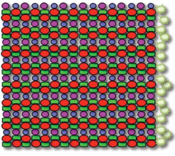
Figure 1.

Schematic of the planar mosaic arrangement of cone photoreceptors in zebrafish: a heterotypic mosaic of cone subtypes organized in a precise, reiterated row pattern. Four cone photoreceptor subtypes are present, including UV (magenta), B (blue), G (green), and R (red) in a precise ratio: twice as many R or G cones relative to UV or B cones, and equal numbers of R and G cones, and equal numbers of B and UV cones. The spatial arrangement is highly stereotyped, with alternating rows of R/G double cones and B/UV single cones. The starbursts represent proliferating cells that give rise to new cone photoreceptors throughout the life of the fish in the marginal germinal zone -- an annulus orthogonal to the cone rows and at the boundary between neural retina and ciliary epithelium.
