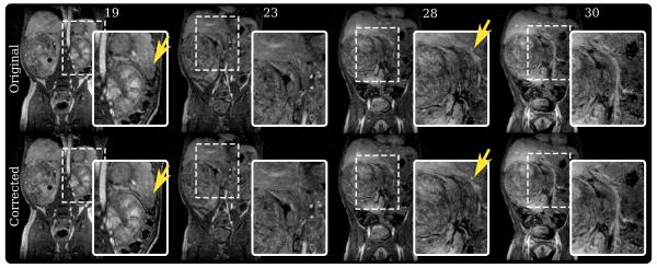
FIG. 7.
Study 2 results – an abdominal study of a 2-year-old patient with a renal tumor scanned using a three-dimensional SPGR sequence. top row: Slice 19, 23, 28, and 30 of the original uncorrected three-dimensional volume. bottom row: Same slices from the corrected volume. Ghosting artifacts in slice 19 were suppressed, and the tissue planes were sharpened. In slice 23, a lesion became better defined after correction.

