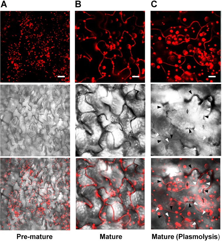
FIGURE 2.
Subcellular localization of PLA2α at different leaf ages in PLA2α-RFP transgenic Arabidopsis plants. (A,B) Epidermal cells in pre-mature leaf tissues (A) and mature leaf tissues (B) from Pro35SPLA2α-RFP transgenic Arabidopsis plants (4-week-old) were viewed using confocal microscopy. (C) Mature leaf tissues were incubated in 1 N KNO3 to induce plasmolysis before being viewed with confocal microscopy. The slightly decreased clarity of images in (C) appears to result from the diffusion of PLA2α-RFP into the expanded apoplasts due to plasmolysis. White arrows in (C) indicate the plasmolyzed plasma membrane, whereas black arrows indicate extracellular spaces. Fluorescent (top), bright field (middle), and merged images (bottom) are presented. Bars = 20 μm.

