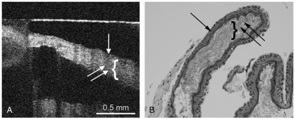
FIG. 3.
A, OCT image of cholesteatoma capsule. Capsule image from the nonkeratinized side, where the cholesteatoma matrix (single arrow) and the keratin (double arrow, bracket) can be observed. B, Histology image of cholesteatoma, where the cholesteatoma matrix (single arrow) and the keratin (double arrow, bracket) can be observed. The horizontal line is an artifact (A).

