
Figure 1. Representative phenomena discussed at the meeting.
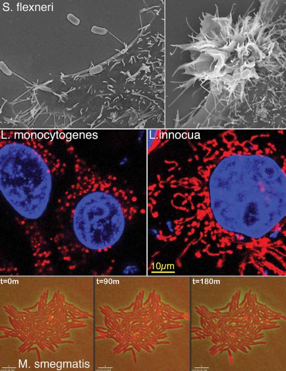
Upper panels, scanning electron micrographs of invasive Shigella flexneri captured by NMEs (left) or inducing membrane ruffling during cell invasion (right); middle panels, fragmentation of mitochondria occurring during Listeria monocytogenes invasion of HeLa epithelial cells (left), which is not observed upon entry of non-pathogenic L. innocua expressing InlB (right). Mitochondria were labelled with cytochrome-C antibody (red) and host cell nuclei and bacteria with Dapi (blue); lower panels, live imaging denoting dynamic expression of a KatG-dsRed fusion protein (red bacteria) in a subpopulation of Mycobacterium smegmatis as a function of time.