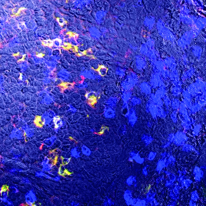
Figure 1.
Immunohistochemistry staining of cervical cancer shows the expression of CD94, NKG2A and CD3 on intratumoral lymphocytes. The stroma tissue at the right side of the picture gives an impression on the high degree of T cell infiltration (CD3 in blue) in cervical cancers. The co-expression of CD94 and NKG2A is predominantly observed on intraepithelial T cells in tumor nests, as can be seen at the left side of the picture. Dr. E. Jordanova is acknowledged for providing this photograph.

