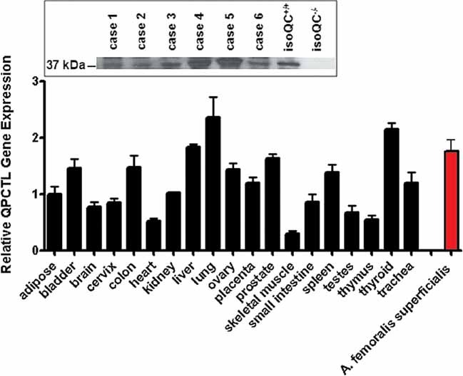
Figure 5. IsoQC is ubiquitously expressed in human tissue.

RT-PCR analysis of various human tissues in comparison to isoQC expression in atherosclerotic vessel segments. The inset shows a Western blot analysis of six cases (5 male, 1 female, mean age 53.8 years) revealing the expression of isoQC in atherosclerotic segments of the Arteria femoralis superficialis compared to expression in livers of isoQC+/+ and isoQC−/− mice as internal standard.
