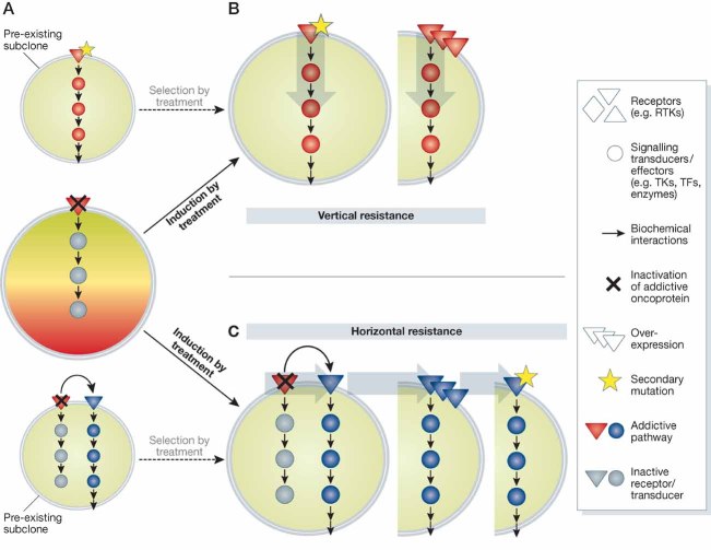
B, C. Fuelled by genomic instability, targeted treatment of addicted cells may result in induction (arrows, solid line) of secondary resistance through either mutation (star) or amplification/overexpression of relevant signalling nodes. This may happen in a vertical fashion, by alterations involving downstream effectors of the original addictive pathway (B), or in a horizontal fashion, when a parallel signalling axis surrogates target blockade (C). If resistant clones are already present at the beginning of treatment (panel A), these may be selected by drug exposure (arrows, dashed line) until they outcompete sensitive cells, resulting in ‘acquired’ resistance as well.

