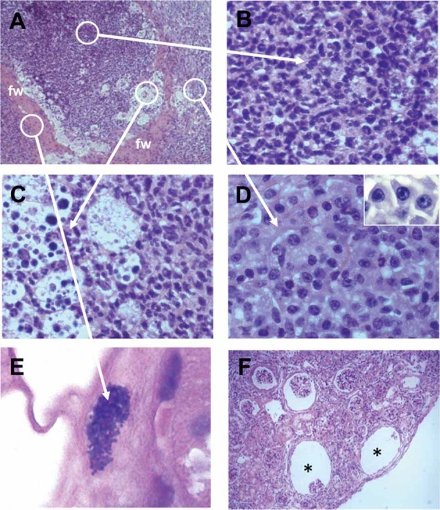
Figure 2. H&E-stained sections of kidney tissue collected from mice at day 56 after S. aureus inoculation.

- S. aureus renal abscess with a peripheral fibrin wall (fw). Original magnification, ×20.
- Cluster of neutrophils occupying the central part of the renal abscess. Original magnification, ×63.
- Rim of apoptotic neutrophils. Original magnification, ×63.
- External layer of B and T cells. Original magnification, ×63. The inset in the upper right corner displays a high magnification (×100) photograph showing cells with the typical morphology of plasma cells.
- Collection of staphylococci can be found associated with fibrin strands. Original magnification, ×100.
- Empty spaces or caverns (asterisks) formed after the resolution of abscesses. Original magnification, ×20.
