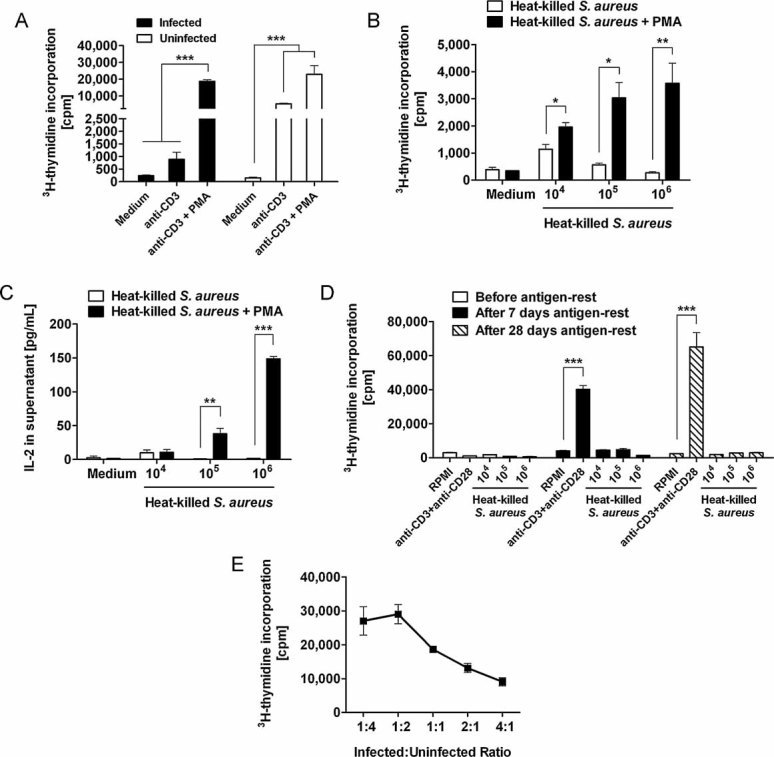
Figure 10. Hyporesponsiveness of T cells to TCR stimulation can be reversed by co-stimulation with PMA.

- Proliferative response of lymphocytes isolated from uninfected (white bars) or from S. aureus-infected (black bars) mice at day 21 of infection to in vitro re-stimulation with anti-CD3 in the presence or absence of the PKC activator PMA. ***p < 0.001.
- Proliferative responses of lymphocytes isolated from S. aureus-infected mice at day 21 of infection to in vitro re-stimulation with increasing concentrations of heat-killed S. aureus in the presence (black bars) or absence (white bars) of PMA. Results are expressed as the mean cpm ± SD as determined from three independent experiments. *p < 0.05 and **p < 0.01.
- Levels of IL-2 in the culture supernatant of spleen cells isolated from S. aureus-infected mice at day 21 of infection and in vitro re-stimulated with increasing concentrations of heat-killed S. aureus in the presence (black bars) or absence (white bars) of PMA. The level of IL-2 was assessed by ELISA and each bar represents the mean value ± SD of three independent experiments. **p < 0.01 and ***p < 0.001.
- Proliferative responses of lymphocytes isolated from S. aureus-infected mice at day 21 of infection to in vitro re-stimulation with increasing concentrations of heat-killed S. aureus or anti-CD3/anti-CD28 before (white bars) or after 7 days (black bars) or 28 days (hatched bars) antigen-rest. Each bar represents the mean value ± SD of three independent experiments. ***p < 0.001.
- Suppressive effect of lymphocytes isolated from S. aureus-infected mice at day 21 of infection in the proliferation of naive T cells. Splenocytes from infected mice were co-cultured with splenocytes from naive mice at different ratios and stimulated with anti-CD3/anti-CD28. Proliferation was measured by 3H-thymidine incorporation, and the data represents the average of three independent experiments.
